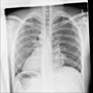
φA medical student is asked to perform a cardiovascular examination on a patient. After 10 minutes of auscultation with no success, the medical student gives up and asks his resident for help. The resident puts up the patient’s anteroposterior chest radiograph (see image) and begins to explain the molecular etiology of this patient’s condition.
(1) Why was the medical student initially overwhelmed??
(2) What is one respiratory complication that this patient is at increased risk of developing as a result of his condition and why??


From the X-ray, it can clearly be seen that the heart is located on the right side of the thorax(dextrocardia). That could explain why the said student was initially overwhelmed, he/she wasn’t getting any reading during the auscultation cus he/she was checking the wrong side.
Hi David, Indeed your attempt is truly remarkable. I especially admire your Keen interest in noting details.
Truly, according to the X-ray film, the heart is placed on the wrong side of the body (right), a condition otherwise called dextrocardia as you rightly noted thus explaining an auscultation with no success by the student.
WHAT IS DEXTROCARDIA?
This is a congenital abnormally in which the heart faces the wrong side (right) of the body. It may be isolated i.e exist alone or associated with other organs or visceral i.e situs invertus. Worthy of note is the fact that this condition is compatible with Life.
In either case (isolated DEXTROCARDIA or Situs Invertus), variants of abnormalities may/may not exist. They include but are not limited to Cardiac abnormalities, Circulatory abnormalities, respiratory abnormalities, etc.
Thank you
Warmest regards
This condition is called dextrocardia in which d heart is facing d right side instead of the left.
It may be an isolated dextrocardia or other forms, the condition can be diagnosed using chest x-ray, CT scan or MRI
The student was overwhelmed because he wasn’t auscultating in the region were d heart is located
The respiratory complication that this patient is at risk is pneumonia and hypoxia(decreased oxygen). The pneumonia can develop as a result of decreased cilia formation within lung tissue, Hence leads to reduced ability of the lungs to clear away microorganism that enter into the lungs.
Dearest Mahmud,
First I must commend your write-up, it is truly impressive. I must also draw your attention to something quite important.
. . .What is Hypoxia?
Hypoxia simply means inability/deficiency of oxygen in tissues. This deficiency may arise as a result of Respiratory complications or variants of other factors not necessarily respiratory in nature. On the basis of this, Hypoxia has been broadly classified into atleast 5subtypes.
Otherthan this Correction, I would say your work was flawless.
Thank you
Warm regards
2. Dextrocardia is More often than none accompanied by a condition known as “Cilliary dyskinesia”.
Cilliary – Cilia.
Dys- Non.
Kinesia- movement.
In this condition, the cilia that helps move the mucus becomes immovable. Which is the respiratory complication of Dextrocardia.
Dearest David,
Ciliary dyskinesia usually accompanies Dextrocardia. As noted by you, the cilia found lining the respiratory pathway is immobile, thus unable to remove mucinous secretions by beating back and forth. These secretions eventually builds up in the Respiratory pathway, clogging as well as expanding the tubes thus leading to probably the most important respiratory complication seen in Dextrocardia known as bronchiectasis.
Thank you.
Warm regards
The condition is termed Dextrocardia ( is the situation why the hearth lies at the right hand side of the chest instead of the left hand side of the chest)..
So the student was over whelmed when he is not hearing the auscultation at the left side not knowing it a defect that the heart lies at the right side.
In shot the respiratory condition of this is that the lungs is supersede by the action of the heart making it difficult for the ciliated muscle if the lung to perform there action…
Hello Virgin,
The response in your first and second paragraph is correct.
In your third paragraph, your responses wasn’t quite clear. kindly clarify us by resending it again.
Thank you
Warm regards
The patient have dextrocardia meaning that the heart due to genetic abnormality is located in the right side of the chest rather than the normal site which is left
The patient has what is termed as kategerner’s syndrome or otherwise called primary ciliary dyskinesia
The syndrome is characterised by the following
Chronic sinusitis
Bronchiectasis
Situs inversus
Dextrocardia
The main respiratory complications that the patient might experience is bronchiectasis
This is so because in the respiratory pathway there are cilia located which are responsible for clearing of mucus along with some particles and from the molecular basis of cilia there are some proteins which helps in the motility of the cilia. So due to the genetic abnormalities of those proteins the cilia is inactive so the clearance of the mucous is impaired which give a way for recurrent respiratory tract infections
So the recurrent respiratory tract infection can leads to bronchiectasis which is among the supparative lung disease
Hi Umar,
This work is both flawless and superb.
No Corrections noted.
Thank you
Warm regards
The above is a classical example of dextrocardia, as observed in the radiograph, the apex of the heart is pointed to towards the right pulmonary cavity.
The student was unable to auscultate this abnormally positioned heart due to the fact that normally the auscultation point for the apex of the heart lies at the 4th to 5th left intercoastal space but in this patient the apex of the heart is directed to the right so no apex heartbeat is auscultated.
(B) The condition if isolated will result in transposition of the great vessels. Here the aorta now opens into right ventricle the pulmonary artery into the left so the systemic supply for left ventricle remains deoxygenated but the pulmonary artery keep carrying oxygenated blood to the lungs, hence the lungs on keep reoxygenating oxygenated blood why the body receives deoxygenated blood resulting in systemic hypoxia, cyanosis, due to abnormal pressure differences pulmonary hypoplasia may set in.
Hi Light,
A great attempt worth commending.
But unfortunately, you weren’t correct in the (B) part of the Question.
Transposition of great vessels (in this case pulmonary artery and aorta) simply means a switch/change in the normal/actual position of both arteries.
Usually the aorta originates from the left ventricle and the pulmonary artery from the right ventricle. In transposition of vessels, the reverse is the case.
I must note then that transposition of great arteries is divided into two subtypes;
(1) Dextro- Transposition of great arteries (TGA)
(2) Levo- Transposition of great arteries (TGA)
In Dextro- Transposition of great arteries, the exact of what you explained occurs i.e cyanosis is present.
. . .But in Levo- Transposition of great arteries, there is absence of cyanosis (acyanosis).
The reason is quite simple, acyanosis occurs because the left ventricle of the heart is found in the right side of the body and the right ventricle of the heart is found on the left side of the body asin DEXTROCARDIA.
Thus even though the arteries are transposed, they still originate from their usual position as seen in DEXTROCARDIA.
Thank you.
Warm regards
The reason the overwhelmed medical student could not hear any heart sounds on the left is because this patient has complete situs inversus. As can be seen from the anteroposterior chest radiograph, the cardiac outline is on the right, as is the gastric bubble. This condition is usually not harmful if the reversal of viscera is complete, but is quite debilitating if the reversal is limited to the heart. Often, situs inversus is associated with Kartagener syndrome, a condition caused by an autosomal recessive defect in the molecular motor protein dynein. This genetic defect results in immotile cilia, impairing a number of important processes. The important findings are: male infertility due to immotile sperm, female infertility due to immotile cilia in the Fallopian tube, recurrent sinusitis due to a failure in bacteria and particle clearance, and bronchiectasis due to a nonfunctional mucociliary elevator